The Treatment Of Inferior Calcaneal Spur
Overview
A heel spur is a buildup of calcium or a bone hook on the heel bone. This is typically the source of most heel pain. It usually takes an X-ray to see the heel spur protruding from the heel. Without proper heel spur treatment, a heel spur cause inflammation and lead to other ailments like plantar fasciitis and Achilles tendonitis. It is important to be examined by an orthopedic specialist.
Causes
Heel spurs are bony outgrowths positioned where the plantar fascia tissue attaches to the heel bone (the calcaneus). Heel spurs seldom cause pain. It is the inflamed tissue surrounding the spur that causes the pain. The Latin meaning of Plantar Fasciitis is, ?Inflammation of Plantar Fascia.? The plantar fascia is a long, thick and very tough band of tissue beneath your foot that provides arch support. It also connects your toes to your heel bone. Each time you take a step, the arch slightly flattens to absorb impact. This band of tissue is normally quite strong and flexible but unfortunately, circumstances such as undue stress, being overweight, getting older or having irregularities in your foot dynamics can lead to unnatural stretching and micro-tearing of the plantar fascia. This causes pain and swelling at the location where the plantar fascia attaches to the heel bone. As the fascia continually pulls at the heel bone, the constant irritation eventually creates a bony growth on the heel. This is called a heel spur.

Symptoms
The Heel Spur itself is not thought to be painful. Patients who experience pain with Plantar Fasciitis are suffering from inflammation and irritation of the plantar fascia. This the primary cause of pain and not the Heel Spur. Heel Spurs form in some patients who have plantar fasciitis, and tend to occur in patients who have had the problem for a prolonged period of time. While about 70 % of patients with plantar fasciitis have a heel spur, X-rays also show about 50 % of patients with no symptoms of plantar fasciitis also have a heel spur.
Diagnosis
Your doctor will discuss your medical history and will examine your foot and heel for any deformities and inflammation (swelling, redness, heat, pain). He/she will analyze your flexibility, stability, and gait (the way you walk). Occasionally an x-ray or blood tests (to rule out diseases or infections) may be requested.
Non Surgical Treatment
A conventional treatment for a heel spur is a steroid injection. This treatment, however, isn?t always effective because of the many structures in the heel, making it a difficult place for an injection. If this treatment goes wrong, it can make the original symptoms even worse. Another interesting means of treatment is Cryoultrasound, an innovative electromedical device that utilizes the combination of two therapeutic techniques: cryotherapy and ultrasound therapy. Treatments with Cryoultrasound accelerate the healing process by interrupting the cycle and pain and spasms. This form of therapy increases blood circulation and cell metabolism; it stimulates toxin elimination and is supposed to speed up recovery.
Surgical Treatment
Have surgery if no other treatments work. Before performing surgery, doctors usually give home treatments and improved footwear about a year to work. When nothing else eases the pain, here's what you need to know about surgical options. Instep plantar fasciotomy. Doctors remove part of the plantar fascia to ease pressure on the nerves in your foot. Endoscopy. This surgery performs the same function as an instep plantar fasciotomy but uses smaller incisions so that you'll heal faster. However, endoscopy has a higher rate of nerve damage, so consider this before you opt for this option. Be prepared to wear a below-the-knee walking cast to ease the pain of surgery and to speed the healing process. These casts, or "boots," usually work better than crutches to speed up your recovery time.
Prevention
If you have not yet developed this condition, you can take steps to protect yourself from it. Most importantly, make it a rule to wear properly fitted footwear. Avoid shoes that have become worn down in the heel, and don't choose shoes that cause you to walk in an abnormal fashion. Maintaining a healthy weight will ensure that undue pressure isn't being put on the ligaments, tendons and bones of your feet. If your job requires a great deal of time on your feet, or if you exercise regularly, be sure to balance periods of activity with periods of rest for your feet.
Physical Rehabilitation For Bursitis Of The Feet
Overview
There is a thick walled fluid filled bursa between the Achilles tendon and the calcaneal bone. The color Doppler images show hypervascularity of the bursal wall. A minimal amount of fluid in the retrocalcaneal bursa can often be found. A retrocalcaneal bursitis is caused by friction of the Achilles tendon over the upper part of the calcaneal bone. It is often an overuse injury found in athletes.
Causes
Repetitive overuse injury of the ankle during long periods of running and or walking. Tight shoes. The heel counter of the shoe constantly rubbing against the back of the heel. Wearing shoes with a low cut heel counter. Abnormal foot mechanics (abnormal pronation). Poor flexibility. Inappropriate training.
Symptoms
Pain when activating the Achilles tendon (running and jumping) and when applying pressure at the point of attachment of the tendon on the heel bone. Contrary to the tenderness occurring with inflammation of the Achilles tendon, the tenderness is localised to the point of attachment to the heel bone.
Diagnosis
Gram stain. A lab test called a Gram stain is used to determine if certain troublesome bacteria are present. Not all bacteria can be identified with a Gram stain, however, so even if the test comes back negative, septic bursitis cannot be completely ruled out. White blood cell count. An elevated number of white blood cells in the bursa's synovial fluid indicates an infection. Glucose levels test. Glucose levels that are significantly lower than normal may indicate infection.
Non Surgical Treatment
Physiotherapy treatment is vital to hasten the healing process, ensure an optimal outcome and reduce the likelihood of injury recurrence in all patients with retrocalcaneal bursitis. Treatment may comprise soft tissue massage (particularly to the calf muscles), joint mobilization (of the ankle, subtalar joint and foot), dry needling, electrotherapy (e.g. ultrasound), stretches, the use of heel wedges, the use of crutches, ice or heat treatment, arch support taping, the use of a compression bandage, exercises to improve strength, flexibility, balance and core stability, education, anti-inflammatory advice, activity modification advice, biomechanical correction (e.g. the use of orthotics), footwear advice, a gradual return to activity program.
Surgical Treatment
Surgery. Though rare, particularly challenging cases of retrocalcaneal bursitis might warrant a bursectomy, in which the troublesome bursa is removed from the back of the ankle. Surgery can be effective, but operating on this boney area can cause complications, such as trouble with skin healing at the incision site. In addition to removing the bursa, a doctor may use the surgery to treat another condition associated with the retrocalcaneal bursitis. For example, a surgeon may remove a sliver of bone from the back of the heel to alter foot mechanics and reduce future friction. Any bone spurs located where the Achilles attaches to the heel may also be removed. Regardless of the conservative treatment that is provided, it is important to wait until all pain and swelling around the back of the heel is gone before resuming activities. This may take several weeks. Once symptoms are gone, a patient may make a gradual return to his or her activity level before their bursitis symptoms began. Returning to activities that cause friction or stress on the bursa before it is healed will likely cause bursitis symptoms to flare up again.
Do Hammer Toes Hurt
 Overview
hammertoes is a painful deformity wherein a toe bends unnaturally and becomes clawlike. This happens because the tendons of the toe contract abnormally, forcing the toe to bend downward and the middle joint of the toe to protrude upward. Although any toe may be affected, hammertoe usually affects the second toe. The toe assumes a clawlike position and cannot be straightened out. When someone with hammertoe wears shoes, the toe is constantly rubbed, so walking may become especially painful if a callus on the sole of the foot or a corn on the top of a toe develops.
Causes
Hammertoe has three main culprits: tight shoes, trauma, and nerve injuries or disorders. When toes are crowded in shoes that are too tight and narrow, they are unable to rest flat, and this curled hammertoes toe position may become permanent even when you aren't wearing shoes due to the tendons of the toe permanently tightening. When the tendons are held in one position for too long, the muscles tighten and eventually become unable to stretch back out. A similar situation may result when tendons are injured due to trauma, such as a stubbed, jammed, or broken toe.
 Symptoms
The most obvious symptoms of this injury will be the the middle toe joint is permanently bent at an angle. In the beginning movement may still be possible but as time passes and the injury worsens the toe will be locked in place and possible require hammer toe correction surgery to fix. Another key indicator of hammer toe is that a lump or corn will form on top of the toe. The toe joint will be painful and walking can cause severe discomfort. Occasionally a callus may form on the sole of the injured foot. If you see any of these symptoms together or have been enduring pain for some time, seeing a podiatrist should be your next step.
Diagnosis
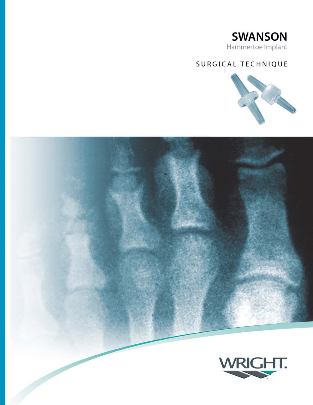
The exam may reveal a toe in which the near bone of the toe (proximal phalanx) is angled upward and the middle bone of the toe points in the opposite direction (plantar flexed). Toes may appear crooked or rotated. The involved joint may be painful when moved, or stiff. There may be areas of thickened skin (corns or calluses) on top of or between the toes, a callus may also be observed at the tip of the affected toe beneath the toenail. An attempt to passively correct the deformity will help elucidate the best treatment option as the examiner determines whether the toe is still flexible or not. It is advisable to assess palpable pulses, since their presence is associated with a good prognosis for healing after surgery. X-rays will demonstrate the contractures of the involved joints, as well as possible arthritic changes and bone enlargements (exostoses, spurs). X-rays of the involved foot are usually performed in a weight-bearing position.
Non Surgical Treatment
Try to find shoes that are soft, roomy, and comfortable and avoid tight shoes or shoes with high heels. A shoe repair shop may be able to stretch a small pocket in regular shoes to make room for the hammertoe. Have a professional pedicure. Sometimes a skilled manicurist can file down a painful corn. Follow your healthcare provider's instructions. Ask your provider what activities you should avoid and when you can return to your normal activities, how to take care of yourself at home, what symptoms or problems you should watch for and what to do if you have them. Make sure you know when you should come back for a checkup.
Surgical Treatment
Surgery to straighten the toe may be needed if an ulcer has formed on either the end or the top surface of the toe. Surgery sometimes involves cutting the tendons that support movement in the toe so that the toe can be straightened. Cutting the tendons, however, takes away the ability to bend the very end of the toe. Another type of surgery combines temporary insertion of a pin or rod into the toe and alteration or repair of the tendons, so that the toe is straightened. After surgery, the deformity rarely recurs.
Hammer Toe Repair Surgery
 Overview
Hammer toes, also called hammer toe, deformity of the second, third, or fourth toe in which the toe is bent downward at the middle joint (the proximal interphalangeal [PIP] joint), such that the overall shape of the toe resembles a hammer. Most cases of hammertoe involve the second toe, and often only one or two toes are affected. In rare cases when all the toes are involved, a thorough neurological assessment is necessary to evaluate for underlying nerve or spinal cord problems.
Causes
As described above, the main reason people develop hammertoes is improper footwear, or footwear that is too short for the toes. Shoes that do not allow our toes to lie flat are the biggest cause of hammertoes, though there are others, including genetics, injury or trauma in which the toe is jammed or broken. Diseases that affect the nerves and muscles, such as arthritis. Abnormal foot mechanics due to nerve or muscle damage, causing an imbalance of the flexor and extensor tendons of the toe. Systematic diseases such as arthritis can also lead to problems such as hammertoe. Some people are born with hammertoes, while others are more prone to developing the condition due to genetics. If you have ever broken a toe, you know there is not much that can be done for it. It is one of the only bones in the body that heals without the use of a cast. A broken toe may be splinted, however, which may help prevent a hammertoe from forming.
 Symptoms
The most common symptoms of hammertoes include. The toe is bent upward at the middle toe Hammer toe joint, so that the top of this joint rubs against the top of the shoe. The remainder of the toe is bent downward. Pain upon pressure at the top of the bent toe from footwear. The formation of corns on the top of the joint. Redness and swelling at the joint contracture. Restricted or painful motion of the toe joint. Pain in the ball of the foot at the base of the affected toe. This occurs because the contracted digit puts pressure on the metatarsal head creating callouse and pressure on the ball of the foot.
Diagnosis
The treatment options vary with the type and severity of each hammer toe, although identifying the deformity early in its development is important to avoid surgery. Your podiatric physician will examine and X-ray the affected area and recommend a treatment plan specific to your condition.
Non Surgical Treatment
Putting padding between your toes and strapping them in place can help to stop pain caused by the toes rubbing. Custom-made insoles for your shoes will help to take the pressure off any painful areas. Special shoes that are wider and deeper than normal can stop your toes rubbing. However if your pain persists your consultant may recommend an surgery.
Surgical Treatment
The technique the surgeon applies during the surgery depends on how much flexibility the person's affected toes still retain. If some flexibility has still been preserved in their affected toes, the hammer toes might be corrected through making a small incision into the toe so the surgeon can manipulate the tendon that is forcing the person's toes into a curved position. If, however, the person's toes have become completely rigid, the surgeon might have to do more than re-aligning the person's tendons. Some pieces of bone may have to be removed so the person's toe has the ability to straighten out. If this is the case, some pins are attached onto the person's foot afterwards to fix their bones into place while the injured tissue heals.
Foot Bunions
Overview
 The best thing to say about the natural history of bunions is that they are unpredictable. This is both in terms of whether the bunion deformity will progress, and also whether the bunion will become painful (if it is not already painful). It would probably however be true to say that once a previously painfree bunion has started to become painful it is not common for the bunion to go back to being entirely pain-free.
Causes
Bunions are caused by a combination of factors, including a familial predisposition, and wearing high-heeled shoes that are tight and narrow at the front. Most bunions occur in women. Sometimes other foot problems accompany bunions, including calluses and hammertoes (angling downward of the lesser toes).
Symptoms
Your bunion may not cause any symptoms. Or you may have pain in your big toe, red or irritated skin over the bunion, and swelling at the base of the big toe. The big toe may point toward the other toes and cause problems in other toes, such as hammer toe . A bunionette can cause similar symptoms at the base of the little toe.
Diagnosis
Your doctor is very likely to be able to diagnose your bunion simply by examining your foot. Even before that, he or she will probably ask about your family and personal medical history and evaluate the types of shoes you wear. You'll be asked about your symptoms, when they started and when they occur. You may also be asked to flex your toe so that your doctor can get an idea of your range of motion. He or she may order x-rays in order to determine the extent of your deformity.
Non Surgical Treatment
Your doctor may recommend a prescription or over-the-counter pain reliever, as well as medication to relieve the swelling and inflammation. A heat pad or warm foot bath may also help relieve the immediate pain and discomfort. A few people may obtain relief with ice packs. If your bunion isn't persistently painful and you take action early on, changing to well-made, well-fitting shoes may be all the treatment you need. Your doctor may advise use of orthoses (devices that are used to improve and realign the bones of your foot), including bunion pads, splints, or other shoe inserts, provided they don't exert pressure elsewhere on the foot and aggravate other foot problems. In some cases, an orthotist (someone trained to provide splints, braces and special footwear to aid movement, correct deformity and relieve discomfort) can recommend shoes with specially designed insoles and uppers that take the pressure off affected joints and help the foot regain its proper shape. 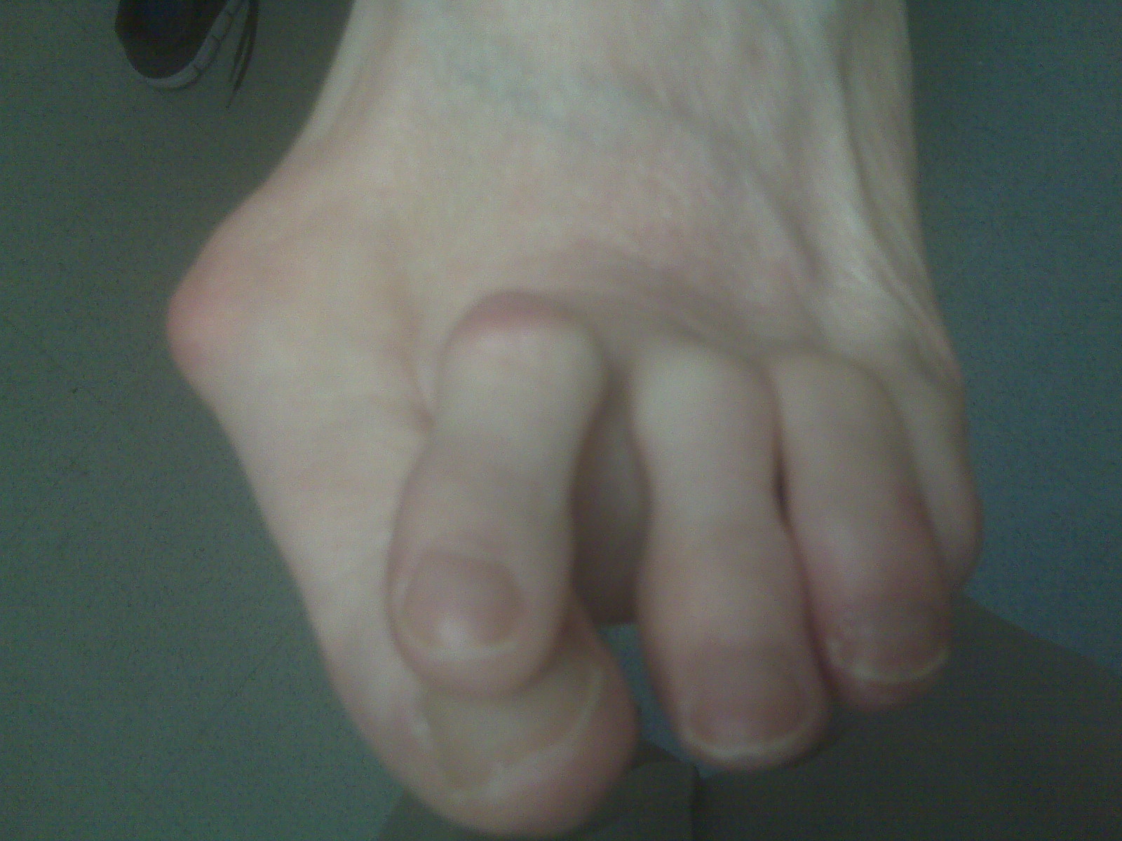
Surgical Treatment
To end persistent pain, your doctor may recommend a type of foot surgery called a bunionectomy to remove the bunion and perhaps to reshape the first toe joint. During the operation, the swollen tissue will be removed, the big toe will be straightened if necessary, and the bones of the affected joint may be reshaped or permanently joined. The goal of the surgery is to correct the cause of the bunion and to prevent the bunion from growing back. After surgery, pain medication will be prescribed, and you will be told when you can start moving your toes and ankle.
Prevention
Bunions often become painful if they are allowed to progress. But not all bunions progress. Many bunion problems can be managed without surgery. In general, bunions that are not painful do not need surgical correction. For this reason, orthopaedic surgeons do not recommend "preventive" surgery for bunions that do not hurt; with proper preventive care, they may never become a problem.
The Facts About Over-Pronation
Overview
Pronation is the inward movement of the foot as it rolls to distribute the force of impact of the ground as you run. The foot "rolls" inward about fifteen percent, comes in complete contact with the ground, and can support your body weight without any problem. Pronation is critical to proper shock absorption, and it helps you push off evenly from the front of the foot. Although pronation is a natural movement of the foot, the size of the arch can affect its ability to roll, causing either supination (underpronation) or overpronation. If you have a normal arch, you're likely a normal pronator, meaning you'll do best in a shoe that offers moderate pronation control. People with flat feet normally overpronate, so they do well in a motion-control shoe that controls pronation. High-arched people typically underpronate, so they do best in a neutral-cushioned shoe that encourages a more natural foot motion.
Causes
Over-pronation is very prominent in people who have flexible, flat feet. The framework of the foot begins to collapse, causing the foot to flatten and adding additional stress to other parts of the foot. Therefore over-pronation can often lead to a case of Plantar Fasciitis, Heel Spurs, Metatarsalgia, Post-tib Tendonitis and/or Bunions. There are many causes of flat feet. Obesity, pregnancy or repetitive pounding on a hard surface can weaken the arch leading to over-pronation. People with flat feet often do not experience discomfort immediately, and some never suffer from any discomfort at all. However, when symptoms develop and become painful, walking becomes awkward and causes increased strain on the feet and calves.
Symptoms
Over-Pronation may cause pain in the heel of the foot, the foot arch, under the ball of the foot, in the ankle, knee, hip or back. The symptoms may be localized to one particular area of the foot or may be experienced in any number of combinations. Standing for long periods of time, walking and running may become difficult due to the additional stress and/or discomfort accompanied with these activities. Upon Visual Inspection, when standing the heels of the foot lean inward and one or both of the knee caps may turn inward.
Diagnosis
So, how can you tell if you have overpronation, or abnormal motion in your feet, and what plantar fasciitis treatment will work to correct it? Look at your feet. While standing, do you clearly see the arch on the inside of your foot? If not, and if the innermost part of your sole touches the floor, then your feet are overpronated. Look at your (running/walking) shoes. If your shoes are more worn on the inside of the sole in particular, then pronation may be a problem for you. Use the wet foot test. Wet your feet and walk along a section of pavement, then look at the footprints you leave behind. A normal foot will leave a print of the heel connected to the forefoot by a strip approximately half the width of the foot on the outside of the sole. If you?re feet are pronated there may be little distinction between the rear and forefoot.
Non Surgical Treatment
Overpronation of the feet can be corrected in some cases and in others it can be effectively managed. Overpronators can train themselves to change their running gait, wear arch supports, orthotic insoles or specialist shoes for overpronators. In order to determine exactly what is happening during the stride, it is necessary to have a gait analysis conducted by a professional. The extent of overpronation can then be determined, and the causes can be identified and corrected directly. The main corrective methods used for excessive pronation are orthotics. Orthotics are the most straightforward and simplest solution to overpronation. Orthotics are devices which can be slipped into shoes which will offer varying degrees of correction to the motion of the foot. Orthotics help to support the arches and distribute the body weight effectively, and are usually the best treatment choice for moderate to severe overpronation. Orthotics may require existing insoles to be removed from your shoes to accommodate them; although most running shoes will have a removable insole to accommodate an orthotic insole.
Prevention
Wear supportive shoes. If we're talking runners you're going to fall in the camp of needing 'motion control' shoes or shoes built for 'moderate' or 'severe' pronators. There are many good brands of shoes out there. Don't just wear these running, the more often the better. Make slow changes. Sudden changes in your training will aggravate your feet more than typical. Make sure you slowly increase your running/walking distance, speed and even how often you go per week. Strengthen your feet. As part of your running/walking warm up or just as part of a nightly routine try a few simple exercises to strengthen your feet, start with just ten of each and slowly add more sets and intensity. Stand facing a mirror and practice raising your arch higher off the ground without lifting your toes. Sit with a towel under your feet, scrunch your toes and try to pull the towel in under your feet. Sitting again with feet on the ground lift your heels as high as you can, then raise and lower on to toe tips.
Calcaneal Apophysitis Physical Therapy
Overview
Sever's disease or calcaneal apophysitis heel pain is a common problem with children between the ages of 8 to 13 years. It has usually been more common in boys, but with the increase of girls in athletic activities, both sexes are having equal symptoms. A high percentage of these children have tight achilles tendons and hamstrings. This condition may occur in the foot with normal arch height or flat or pronated foot, but can be especially painful in the high arch foot.
Causes
The more active a child is then the greater the chance of suffering from Sever?s disease. Poor foot function such as flat feet causes the calf and Achilles to work harder and pull on the growth plate leading to Sever?s disease. Tight calves or Achilles is common in growing children and can increase tension on the growth plate.
Symptoms
The most obvious sign of Sever's disease is pain or tenderness in one or both heels, usually at the back. The pain also might extend to the sides and bottom of the heel, ending near the arch of the foot. A child also may have these related problems, swelling and redness in the heel, difficulty walking, discomfort or stiffness in the feet upon awaking, discomfort when the heel is squeezed on both sides, an unusual walk, such as walking with a limp or on tiptoes to avoid putting pressure on the heel. Symptoms are usually worse during or after activity and get better with rest.
Diagnosis
To diagnose the cause of the child?s heel pain and rule out other more serious conditions, the foot and ankle surgeon obtains a thorough medical history and asks questions about recent activities. The surgeon will also examine the child?s foot and leg. X-rays are often used to evaluate the condition. Other advanced imaging studies and laboratory tests may also be ordered.
Non Surgical Treatment
See a doctor, who can diagnose the injury and recommend appropriate treatment options. It will be beneficial to rest the affected heel, and to regularly ice the affected area for the first few days. Anti-inflammatory pain medication can reduce pain and swelling, but first check with your doctor. As the pain diminishes a physical therapist can assist with a program of rehabilitation, incorporating stretching and strengthening exercises focused on the calf, shin and hamstring muscles. For a period after the injury has healed the doctor may advise on changes to your training routines that seek to lessen the strain on the heels. Orthotics are often recommended for your shoes in order to correct any biomechanical problems or lend extra support to the heels.
Prevention
Sever's disease may be prevented by maintaining good joint and muscle flexibility in the years leading up to, and during, their growth spurts (eg girls 8 to 10, boys 10 to 12). Foot arch problems such as flat feet should be addressed after the age of five if they don't appear to be self-correcting. If you are concerned, please ask your health practitioner. The most important factor is the amount of weight-bearing exercise your child is currently performing.